
Accueil > Séminaires > Année 2023 > Séminaire d’Alexandra Fragola (10 janvier)
Séminaire d’Alexandra Fragola (10 janvier)
par - 2 décembre 2022 (modifié le 3 janvier 2023)
Le séminaire aura lieu dans l’amphi du bât 520 et sera également diffusé en visioconférence. Les personnes extérieures au laboratoire qui souhaitent disposer du lien sont invitées à envoyer un mail à l’adresse
seminaires.ismo@universite-paris-saclay.fr
Elles recevront le lien pour se connecter en retour.
Structured illumination and adaptive optics fluorescence microscopy for high resolution bio-imaging
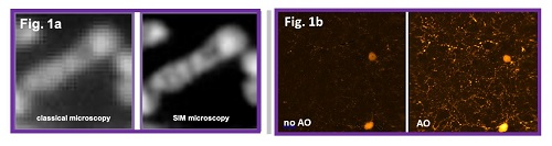
Fluorescence microscopy is a technique of choice for dynamic imaging of living samples. However, image quality remains limited by diffraction or optical aberrations, which restrict both resolution and sensitivity in all microscopy modalities. To overcome these difficulties, we developed new optical microscopes for high-resolution imaging of living biological samples, from the individual cell to the small animal. Structured illumination microscopy (SIM) allows to exceed the diffraction limit with only a few acquired images, thus allowing dynamic super resolution imaging of living cells. As shown in figure 1a, the resolution gain of SIM up to 100nm allows to study the dynamics of the mitochondrial cristae in cells subjected to a photoinduced stress. For in depth imaging inside biological tissues, adaptive optics provides a reliable live correction of the aberrations and thus a significant gain in both spatial resolution and signal intensity. We proposed a few years ago an innovative strategy of adaptive optics, by adapting pioneer work from astronomy to the constraints of fluorescence light-sheet microscopy and two-photon microscopy, and demonstrated its efficiency for enhanced live imaging of Zebrafish larvae and structural or functional neuroimaging of ex vivo Drosophila brains and mouse brain slices (see figure 1b).
- Fig. 1
- 1 (a) Classical and SIM microscopy images of HeLa cells mitochondria labeled with MTG (b) 2-photon fluorescence images without and with adaptive optics of a GADGFP mouse brain slice
Dans la même rubrique :
- Robin Corgier (19 déc)
- d’INTERET GENERAL de Matteo Cacciari (12 déc)
- Philippe-Henri Secrétan et Bernard Do (15 nov)
- d’An Tran (28 nov)
- Sotirios Papadopoulos (14 nov)
- Dennis Tokaryk (10 nov)
- Lucile Rutkowski (7 nov)
- Miriam Kappe (17 oct)
- Kazuki Sumida (4 oct)
- Taichi Okuda (2 oct)
- Freek Massee (28 mars)
- FFJ : Sarah Abrahamsson (21 mars)
- Thomas Pons (14 février)
- Raphaël Hahn (30 janvier)
- Benoît Darquié (24 janvier)
- d’Oliver S. Wenger (ANNULE)
- Ralph Püttner (4 avril)
- d’Audrey Scognamiglio
- Tatiana Itina (11 juillet à 14h)
- P. Bryan Changala (12 septembre)
- d’Ingo Fischer (3 octobre)