Below, you will find some of our research topics. The common factor is the excitation/ionization of species (atoms/molecules/ions) by one or more photons. Our research focuses specifically in the detection of the resulting charged photoproducts (ions/electrons) from which we can infer the mechanism of interest.
This research can be decomposed into:
- Time-resolved studies: we use a combination of ultrafast laser pulses (1 fs = 10-15s) to pump the system of interest and probe it at a later time. By scanning the delay between the two pulses, we can map the dynamics in “real-time”, essentially making a movie.
- Spectrally-resolved studies: This is based on using photon with a well defined energy to preciselly access specific energy levels.
Helium nanodroplets

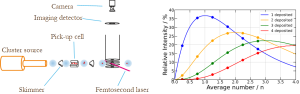
Rare gas clusters offer an elegant approach to capture species, such as atoms and molecules, and rapidly cool to the cluster temperature, enabling formation of weakly-bound complexes usually unattainable in gas-phase experiments. A key advantage of this method is the precise control over stoichiometry, allowing selective formation of specific complexes (b).
Among rare gas aggregates, helium droplets (a) provide a transparent “interaction-free” environment with a sub-Kelvin temperature of 0.37K. They remain transparent below 19.82 eV ensuring only embedded species interact with the light field during using valence shell excitations/ionizations.
Dopant behavior in HeDs is governed by helium affinity. Most molecular species are heliophilic and reside in the center of the droplet with low thermal energy (d1). In contrast, alkali metals and some open-shell molecules exhibit heliophobic character, residing on the droplet surface (d2). Multiple trapped species typically interact to form stable complexes in shallow potential energy wells. Excess binding energy dissipates via He atoms evaporation (b), effectively stabilizing newly formed complexes (1 He atom dissipates about ~0.6 meV). These characteristics have led some researchers to describe HeDs as “flying nano-cryo-reactors” or more modestly “ultimate spectroscopy matrices” due to their unmatched ability to host and probe reactive species with minimal perturbation.
In our laboratory, we use Helium droplet source in conjunction with ultrafast laser pulses to study a broad range of dynamics, starting from reaction dynamics between two isolated species, solvent relaxation, or gas-phase like experiments with ultracold molecules.
Time-resolved reaction dynamics
Time-Resolved Reaction Dynamics (TRRD) emerged with the development of ultrashort laser pulses and first aimed at addressing the very fundamental question: “Is it possible to follow a bimolecular reaction in real time all the way from the reactants to the products?”. The reactants and products can usually be characterized by frequency-resolved spectroscopy, but following the reaction in real time requires time-resolved methods with sufficient time resolution. TRRD focuses on tracking how chemical bonds break and form. More precisely, this field concentrates on the comprehension, the characterization and the manipulation of the forces that drive the chemical reaction. These studies are thus overcoming the static picture of bonds to project it entirely onto the dynamical aspect of the electronic and oncoming nuclear dynamics, often out of the Born-Oppenheimer approximation.
In this context, one aspect of the research focuses on the reaction dynamics between two species (atoms/molecules) embedded in a superfluid environment, hereby named Helium nanodroplet.Using a combination of ultrafast of laser pulses, we can induce and track in real time the reaction between the two species by exciting/ionizing one of them.
Relaxation dynamics
The photoexcitation of a molecule can lead to a large amount of internal energy during its relaxation through different electronic states. This relaxation typically involves numerous degrees of freedom and may lead to its fragmentation or isomerisation. In any case, this internal energy is usually a drawback as it leads to a blurring of some observables.
Consequentlty, in the case of isomerization, the different photoproducts cannot be identified properly as their photoelectron spectra overlap. This effect is well known and is usually defined as spectral congestion.
One way to circumvent this limitation, is to do the experiments in a solvent. In that case, the internal energy can be dissipated through the solvent excitation. In the case of HeDs, the low temperature of 0.37K permits to differentiate the photoproducts and also allow to preserve the molecules from fragmentation.
The dynamics of a chemical reaction is an essential information that must be obtained to obtain a detailed vision of the mechanism of that reaction. The chemical phenomenon studied is photoinduced by a laser. This excitation occurs without geometric change of the reactive system, but the potential gradients in the excited state give the system the necessary impetus for the reaction. Temporal information allows for the exploration of the passage of the reactive system through geometric conformations and electronic configurations that are very distant from those accessible in the Franck-Condon region.
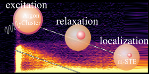
Exciton relaxation in clusters and isolated nanoparticles
We combined time-resolved photoelectron spectroscopy on isolated species with high-level data processing to address a question that generally falls under materials science: the dynamics of electronic relaxation towards the formation of a self-trapped exciton (STE). These excitons are common excited states in ionic crystals, silica, and rare gas matrices. They are associated with a strong local distortion of the matrix. Argon aggregates were taken as a model system. They are initially excited to a Wannier exciton at 14 eV, and their evolution towards the formation of an STE exhibited an unusual type of vibronic relaxation where the electronic excitation of the aggregate decreases linearly over time at a rate of 0.59 eV/ps. The decay was monitored for 3.0 ps, and the formation of STE occurred in about 5.1 ps.
Cluster Isolated Chemical Reactions (CICR)
Studying chemical reactions in an aggregate is an elegant way to investigate the effects of a reaction medium on the dynamics of a chemical reaction. The “Cluster Isolated Chemical Reaction” (CICR) technique is particularly interesting in this field due to its flexibility: this technique allows for the selection of a known number of reactive atoms or molecules and their interaction with a medium, the aggregate, whose structure and temperature are known. The aggregate plays two successive roles: first, it serves to collisionally capture the reactants, and then it mimics the solvent. The motivations for isolating reactions on van der Waals (or helium) aggregates are of several types:
Using the flexibility of the CICR technique to create a study object that is difficult to obtain otherwise, while ensuring spectroscopic information about it.
Determining the exact stoichiometry of a reaction.
Studying how the aggregate disrupts the reaction occurring within it. Two effects exist: i) mechanical by hindering movements, ii) electronic by altering the relative heights of the different potential surfaces involved in the studied chemical reactions, or even modifying the surfaces themselves. The induced dipole/permanent dipole interaction indeed stabilizes the potential surfaces or the regions of potential surfaces corresponding to a charge transfer compared to those for which there is no charge transfer.
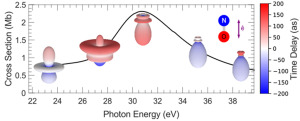
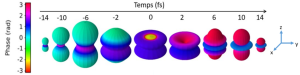
Attosecond photoionization delays
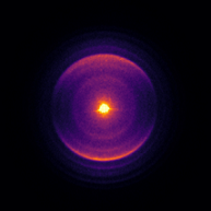
The time delay of a photoelectron emitted angularly resolved in the molecular reference frame carries the signature of the interaction of the electron ejected from a molecular orbital by the absorption of an XUV photon with the electrostatic potential representing the electronic structure of the molecule. The determination of this observable is generally the objective of complex two-photon (XUV – IR) experiments, temporally resolved on the attosecond scale. We have demonstrated that such photoionization delays can be determined by measuring the molecular emission diagrams of photoelectrons spectrally resolved in the XUV domain using synchrotron radiation (SOLEIL). The reaction illustrating these experimental and theoretical results involves the interference between direct ionization and resonant ionization due to a shape resonance, described by a multichannel Fano model (see figure). The resonant state, responsible for the modulation of the cross-section, increases the ionization delay by retaining the electron before ionization and modifies the emission diagram.
The ionization time delay can also be measured using a time-resolved approach, employing the RABBIT method which involves measuring the time difference taken by an electron escaping from an ion, for different ionization energies. In collaboration with LIDYL, ILM, and LCPMR, and by introducing an angular approach to the measurement, we were able to reconstruct the film of the ionization process in helium.
Isolated nanoparticle-femtosecond laser interaction
I am text block. Click edit button to change this text. Lorem ipsum dolor sit amet, consectetur adipiscing elit. Ut elit tellus, luctus nec ullamcorper mattis, pulvinar dapibus leo.
Ion spectroscopy
The study of photoionization processes in ionic species is essential for the characterization and modeling of many plasmas, whether they are laboratory-based (laser plasmas, tokamaks, etc.) or of astrophysical interest (stars, nebulae, etc.) and planetary (ionospheres). From a fundamental perspective, understanding photon-matter interactions in complex atomic systems poses a real challenge for theoretical models aimed at describing the various competing interactions within the atomic electronic structure. The photoionization process allows, among other things, for tracking the evolution of the relative intensity of electronic correlations in isonuclear series as a function of the variation in the intensity of the central potential.
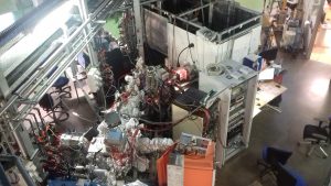
Experiments on the photoionization of ionic species began in the 1980s using synchrotron radiation provided by the ACO ring and continued at Super ACO, and then on the Pléiades beamline at Soleil, where the current MAIA experimental setup has a permanent installation. Only one setup, located in Germany, competes with it.
Among the numerous results obtained, we can mention the study of the collapse of the 4f orbital in iodine, xenon, or barium depending on the initial charge state of the ion, the study of isonuclear series of ions of astrophysical interest (C, N, O, Fe), or the study of heavy systems whose modeling requires the use of UTA. These studies have extended to more complex systems such as hydrides.
Collaborations:
E.T. Kennedy, J.P. Mosnier (Dublin College University)
C. Blancard (CEA Bruyères-le-Châtel)
B. Mc Laughlin (Harvard Smithsonian Center for Astrophysics)
T. Gorczyca (Western Michigan University)
S. Cargniato (LCPMR, Paris)
Velocity Map Imaging Spectrometers (VMIS)
I am text block. Click edit button to change this text. Lorem ipsum dolor sit amet, consectetur adipiscing elit. Ut elit tellus, luctus nec ullamcorper mattis, pulvinar dapibus leo.
Femto and Attosecond XUV pulses
I am text block. Click edit button to change this text. Lorem ipsum dolor sit amet, consectetur adipiscing elit. Ut elit tellus, luctus nec ullamcorper mattis, pulvinar dapibus leo.